

CAE Blue Phantom™ Gen II Ultrasound Central Line Model With Transparent Insert & Hand Pump
Training model with transparent tissue insert allows users to develop and practice the skills necessary to gain proficiency in using ultrasound to guide central catheter insertions in the internal jugular vein (IJ), subclavian vein, and axillary vein
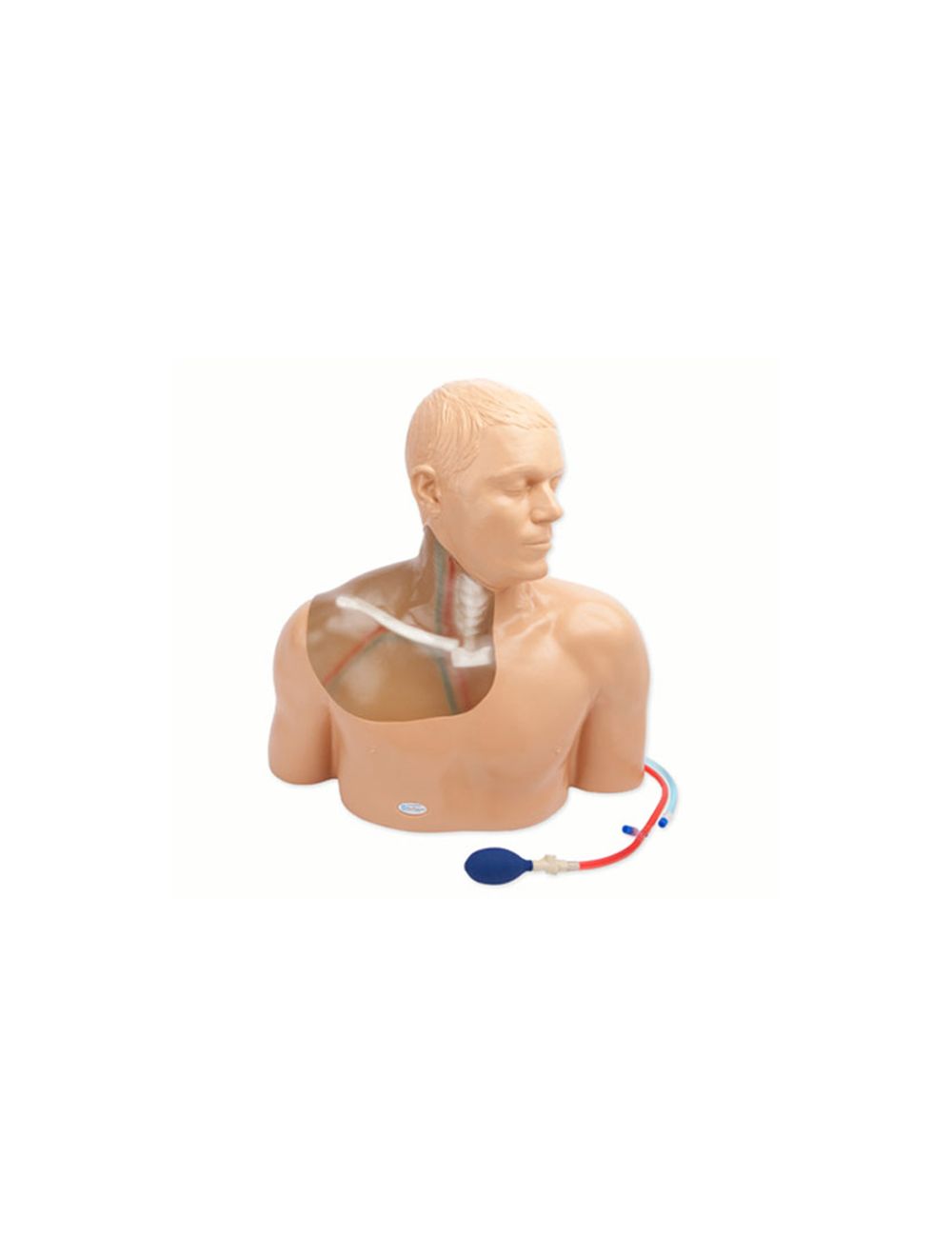
Second Generation upper torso ultrasound guided central line placement training model with transparent tissue insert allows users to develop and practice the skills necessary to gain proficiency in using ultrasound to guide central catheter insertions in the internal jugular vein (IJ), subclavian vein, and axillary vein while revealing the venous and arterial vessels as well as accessory boney structures. Developed with the goal of helping clinicians bridge the learning gap by allowing them to see the internal anatomical structures with their eyes as well as with ultrasound imaging, our transparent central line placement tissue offers superb ultrasound hands-on training. Using Blue Phantom proprietary simulated human tissue, this very realistic and ultra-durable transparent central venous access ultrasound training model is excellent for training clinicians in the psychomotor skills associated with ultrasound guided central line placement procedures. These ultrasound imaging skills include; using ultrasound system controls, transducer positioning and movement, recognition of arterial and venous anatomy, using ultrasound to target the appropriate vessels for cannulation, and performing a central venous access procedure.
Each model offers anatomically correct vascular anatomy of the right upper thorax and neck including the internal jugular vein, brachiocephalic vein, subclavian vein, axillary vein, carotid artery, subclavian artery, and axillary artery, as well as anatomical landmarks including the clavicle, the two heads of the sternocleidomastoid muscle, and the sternal notch. Users can utilize traditional anatomical landmarks for blind insertion techniques, or utilize ultrasound to obtain images of anatomical structures. A simulated SVC accommodates full threading of guidewires, dilators and central line catheter placement. Venous and arterial fluids that are removed during central catheter insertions training are easily refilled using quick fill ports. Users can run intravenous fluid for constant refill of vessels during training.
Arterial pulsations can help users differentiate between arteries and veins. Three arterial pulse configurations are available; no pump, hand pump, and integrated automated pump. Points of access include IJ, subclavian, infraclavicular and supraclavicular approach as well as access via the axillary vessel. Positive fluid flow in the vessels provides users with immediate feedback when vessels are accessed. The simulated blood fluids in the arterial vessels differ from the venous system allowing for users to easily differentiate between successful and unsuccessful venous access procedures.
- Manufactured using Blue Phantom patented ultra-durable tissue and is extremely realistic in ultrasound imaging characteristics feels and cannulates like real human tissue
- Realistic and ultra-durable central venous access ultrasound training model excellent for training clinicians in the psychomotor skills associated with ultrasound guided central venous access procedures
- Developed with the goal of helping clinicians bridge the learning gap by allowing them to see the internal anatomical structures with their eyes as well as with ultrasound imaging
- Superb ultrasound imaging characteristics
- Ultra-durable self healing tissue is extremely realistic in ultrasound imaging characteristics and feels like real human tissue
- Self healing tissue will withstand tremendous use and will save you money by dramatically reducing the necessity for purchasing replacement parts
- Contains anatomically correct vascular anatomy of the right upper thorax and neck including the internal jugular vein, brachiocephalic vein, subclavian vein, axillary vein, carotid artery, subclavian artery, and axillary artery, as well as anatomical landmarks including the clavicle, the two heads of the sternocleidomastoid muscle, and the sternal notch
- Accommodates full threading of guidewires and catheters
- Venous and arterial fluids that are removed during central catheter insertions training are easily refilled using quick fill ports
- Three arterial pulse configurations are available; no pump, hand pump, and integrated automated pump
- Positive fluid flow in the vessels provides users with immediate feedback when vessels are accessed
- Simulated blood fluids in the arterial vessels differ from the venous system allowing for users to easily verify successful venous access procedures
- Tissues match the acoustic characteristics of real human tissue so when you use your ultrasound system on our training models, you experience the same quality you expect from imaging patients in a clinical environment
- Performs well using any ultrasound imaging system
- Practice using ultrasound system controls
- High quality
- No special storage needs
- Patented technology
Includes:
- 2 bottle of simulated refill solution; one red (arterial), one blue (venous). 235mL bottles.
Dimensions: 17” x 11” x 17” (43cm x 28cm x 43cm) (L x W x H)
Weight: 20 lbs. (9 Kg.)
Made in USA
EXCEPTIONAL
CUSTOMER SERVICE
PRODUCTS YOU NEED
PRICES YOU'LL LOVE
SPEEDY SHIPPING
WORLDWIDE