

Erler Zimmer Small Arterial Leg Ulcer Wound
Moulage wounds of different wound stages of arterial leg ulcers
Options by Part Number:
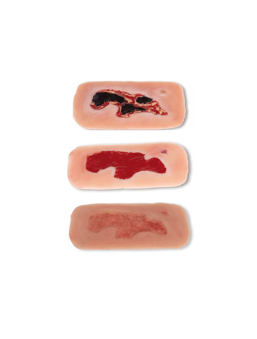
53-134 Small Arterial Leg Ulcer in Exudation Phase Wound
53-135 Small Arterial Leg Ulcer in Granulation Phase Wound
53-136 Small Arterial Leg Ulcer in Epithelialization Phase Wound
These self-adhesive wounds represent arterial leg ulcers, substance defects in the tissue of the lower leg as a result of a peripheral arterial occlusive disease (PAOD). Caused by insufficient blood and related oxygen supply the healing of wounds is inhibited any small injuries can become chronic wounds. These small wounds are perfect for adhering at the ankle or lower leg.
The moulage adheres independently to standardized patients and manikins. For longer training or improved adherence, an adhesive (sold separately) may be used.
- Exudation Phase: Necrosis of tissue is represented as extensive black areas.
- Granulation Phase: the granulation phase after successful causal therapy and careful wound cleaning is represented
- Epithelialization Phase: shows the building of epithelial tissue, finally leading to closure of the wound after successful causal therapy and careful wound cleaning
Individual wounds include:
- Storage case
2-year warranty
EXCEPTIONAL
CUSTOMER SERVICE
PRODUCTS YOU NEED
PRICES YOU'LL LOVE
SPEEDY SHIPPING
WORLDWIDE